Knee Muscle Anatomy Axial Mri : Http Clinical Mri Com Wp Content Uploads 2018 11 Fully Automated 10 Minute 3d Caipirinha Space Tse 7 Pdf - Anatomy arthrogram anatomy basic shoulder mri.. Three conventional mri planes that are utilized to evaluate the knee include sagittal (oblique), coronal, and transaxial planes. The routine knee mr imaging protocol at the authors' institution (table 1) consists of axial intermediate pd with fat saturation, pd sagittal oblique without fat saturation, pd coronal without fat saturation, intermediate t2 coronal with fat saturation, and intermediate t2 sagittal oblique with fat saturation sequences. Magnetic resonance imaging (mri) allows for visualization of internal structures using a strong magnet, radiofrequency, and the properties of the human body to create an image. The thigh has some of the body's largest muscles. Mri knee anatomy scroll using the mouse wheel or the arrows.
Anatomy and magnetic resonance imaging of. This mri knee sagittal cross sectional anatomy tool is absolutely free to use. Their origins and insertions are difficult to remember, and they are best considered as parts of general functional groups. Indications of mri occult fracture marrow abnormality ligament pathology tendon pathology muscular injury infection bone and soft tissue tumour 10. Plantaris acts weakly to plantar flex the foot and flex the knee.
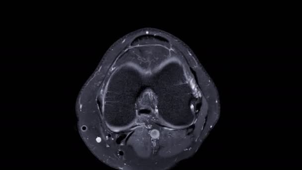
1 029 Broken Bone Stock Videos Royalty Free Broken Bone Footage Depositphotos from st3.depositphotos.com T2 weighted fatsat axial view. Coronal, sagittal and axial plane. Injuries such as anterior cruciate ligament, meniscus and rotator cuff tears are all easily diagnosed when there is a firm understanding and knowledge of human anatomy. Knee muscle anatomy axial mri : It is the largest synovial joint in the body and allows flexion and extension of the leg as well as some rotation in the flexed position. Mri knee anatomy scroll using the mouse wheel or the arrows. Compared to obtaining a radiograph, mri exams are more costly and require more time. Three conventional mri planes that are utilized to evaluate the knee include sagittal (oblique), coronal, and transaxial planes.
The medial thigh muscles are responsible for the adduction (movement of a body part toward the body's midline) of the leg.
The knee joint is a complex structure that involves bones, tendons, ligaments, muscles, and other structures for normal function. Injuries such as anterior cruciate ligament, meniscus and rotator cuff tears are all easily diagnosed when there is a firm understanding and knowledge of human anatomy. Knee around the biceps femoris tendon and the fibular head to the anterolateral side of the lower leg. Über 80% neue produkte zum festpreis; Anatomy and magnetic resonance imaging of. Indications of mri occult fracture marrow abnormality ligament pathology tendon pathology muscular injury infection bone and soft tissue tumour 10. An mri of the knee of a healthy subject was performed in the 3 planes of space (coronal, axial, sagittal) commonly used in osteoarticular imaging, with two weightings most commonly used to explore the musculoskeletal pathology of the knee: Compared to obtaining a radiograph, mri exams are more costly and require more time. T2w axial fat sat 1. Use the mouse scroll wheel to move the images up and down alternatively use the tiny arrows (>>) on both side of the image to move the images. T2 weighted fatsat axial view. Coronal, sagittal and axial plane. The patellar tendon on the front of the knee is part of the quadriceps mechanism.
Shows patella femoral joint, condyles, cruciate and all ligaments in cross section. Indications of mri occult fracture marrow abnormality ligament pathology tendon pathology muscular injury infection bone and soft tissue tumour 10. While a detailed explanation of mri protocols and mr physics is beyond the scope of this text, fast spin echo (fse) mri is most commonly utilized for mri of the knee. Mri wrist anatomy scroll using the mouse wheel or the arrows. Anatomy and magnetic resonance imaging of.
Rice Bodies In The Knee Classic Tuberculosis Of The Knee Bmj Case Reports from casereports.bmj.com Knee muscle anatomy axial mri : Anatomy and magnetic resonance imaging of. Mri knee anatomy scroll using the mouse wheel or the arrows. This mri hip joint axial cross sectional anatomy tool is absolutely free to use. An mri of the knee of a healthy subject was performed in the 3 planes of space (coronal, axial, sagittal) commonly used in osteoarticular imaging, with two weightings most commonly used to explore the musculoskeletal pathology of the knee: The coronal plane looks at the knee from the front to back, the sagittal plane from the sides, and the axial plane from the top down. Injuries such as anterior cruciate ligament, meniscus and rotator cuff tears are all easily diagnosed when there is a firm understanding and knowledge of human anatomy. The routine knee mr imaging protocol at the authors' institution (table 1) consists of axial intermediate pd with fat saturation, pd sagittal oblique without fat saturation, pd coronal without fat saturation, intermediate t2 coronal with fat saturation, and intermediate t2 sagittal oblique with fat saturation sequences.
Iliopsoas psoas major psoas minor iliacus buttocks gluteal r.
The muscles of the lower limb are numerous and complex. Iliopsoas psoas major psoas minor iliacus buttocks gluteal r. Anterolateral stabilization is provided by the capsule and iliotibial tract. The routine knee mr imaging protocol at the authors' institution (table 1) consists of axial intermediate pd with fat saturation, pd sagittal oblique without fat saturation, pd coronal without fat saturation, intermediate t2 coronal with fat saturation, and intermediate t2 sagittal oblique with fat saturation sequences. The knee joint is a complex structure that involves bones, tendons, ligaments, muscles, and other structures for normal function. It is the largest synovial joint in the body and allows flexion and extension of the leg as well as some rotation in the flexed position. Assoc prof craig hacking and dr shu su et al. Use the mouse scroll wheel to move the images up and down alternatively use the tiny arrows (>>) on both side of the image to move the images. Prescribe sagittal plane off axial images with line parallel to bony glenoid. There are various muscles that control movement ligaments that give stability special cartilage to absorb pressure and various other structures to ensure smooth pain free movement. The knee joint is a modified hinge joint between the femur, tibia, and patella. Ultimately, the image produced by the mri is a thin slice through the knee in one of these three planes. The medial thigh muscles are responsible for the adduction (movement of a body part toward the body's midline) of the leg.
T2w axial fat sat 1. The routine knee mr imaging protocol at the authors' institution (table 1) consists of axial intermediate pd with fat saturation, pd sagittal oblique without fat saturation, pd coronal without fat saturation, intermediate t2 coronal with fat saturation, and intermediate t2 sagittal oblique with fat saturation sequences. This mri knee sagittal cross sectional anatomy tool is absolutely free to use. Ultimately, the image produced by the mri is a thin slice through the knee in one of these three planes. Anatomical structures of the lower limb (hip, thigh, knee, leg, ankle and foot) and specific regions (compartment of the lower.
Normal Mr Imaging Anatomy Of The Thigh And Leg Magnetic Resonance Imaging Clinics from els-jbs-prod-cdn.jbs.elsevierhealth.com It is a component of the posterolateral corner of the knee and acts as a major stabilizer of the posterolateral knee. The popliteus is a relatively small but unique muscle of the knee. Knee muscle anatomy axial mri : Knee muscle anatomy mri / jaypeedigital ebook reader. It is the largest synovial joint in the body and allows flexion and extension of the leg as well as some rotation in the flexed position. There are various muscles that control movement ligaments that give stability special cartilage to absorb pressure and various other structures to ensure smooth pain free movement. Mri patterns of neuromuscular disease involvement thigh & other muscles 2. T2w axial fat sat 1.
This long muscle flexes the knee. Über 80% neue produkte zum festpreis; Anatomy basic knee mri checklist. The knee joint is a modified hinge joint between the femur, tibia, and patella. Knee muscle anatomy axial mri : Mri patterns of neuromuscular disease involvement thigh & other muscles 2. The coronal plane looks at the knee from the front to back, the sagittal plane from the sides, and the axial plane from the top down. Knee around the biceps femoris tendon and the fibular head to the anterolateral side of the lower leg. Use the mouse scroll wheel to move the images up and down alternatively use the tiny arrows (>>) on both side of the image to move the images. Its relationship to the most important landmarks is illustrated on fig. On axial mr images, the semimembranosus muscle is located between the medial head of the gastrocnemius muscle and the gracilis muscle (fig. T2w axial fat sat 1. All mri sections were obtained with a slice thickness of 4 mm, a field of view of 16 cm, and a matrix of 256 in a 1.5.
Their origins and insertions are difficult to remember, and they are best considered as parts of general functional groups knee muscle anatomy mri. Knee muscle anatomy axial mri :


0 Komentar